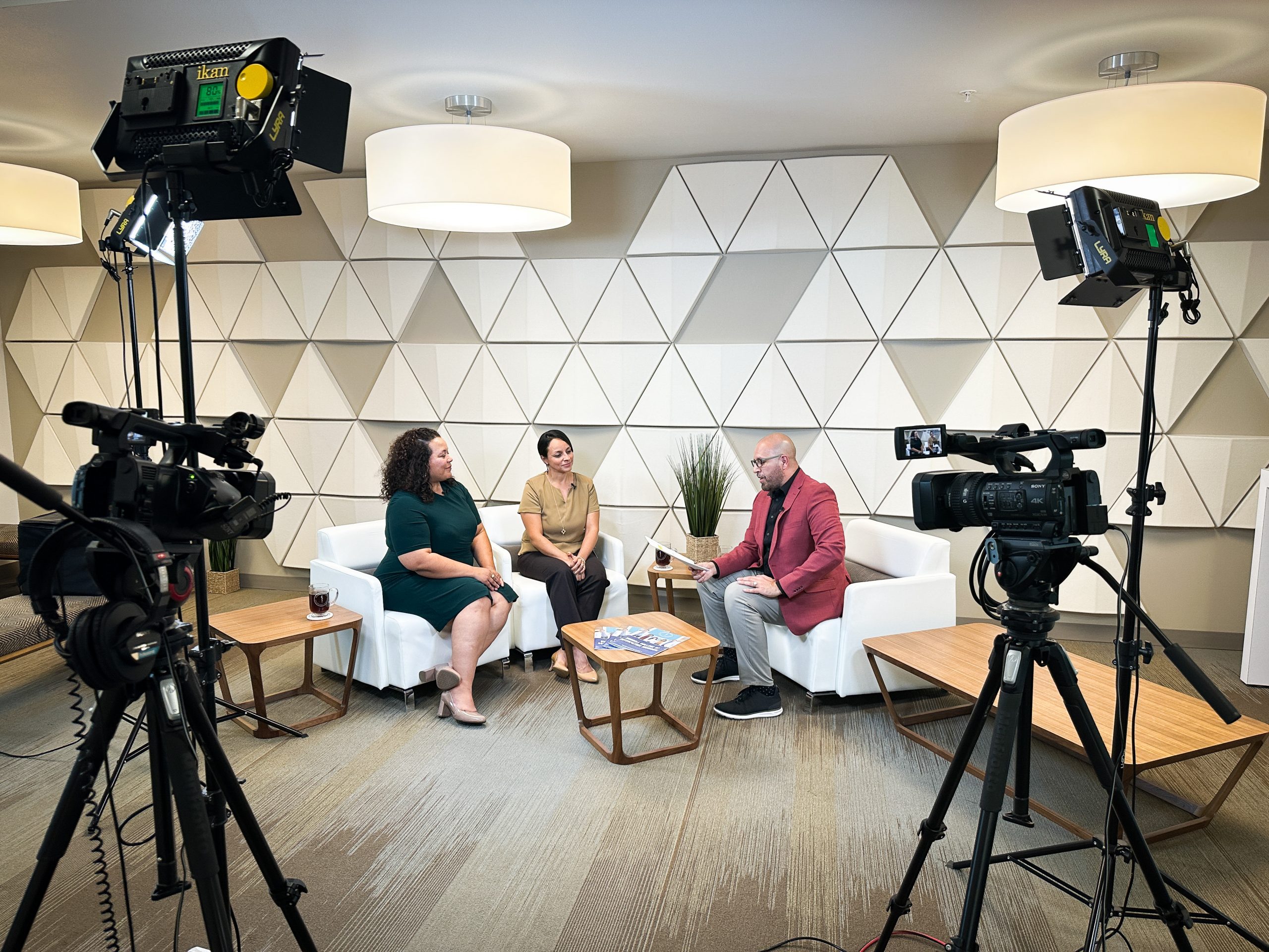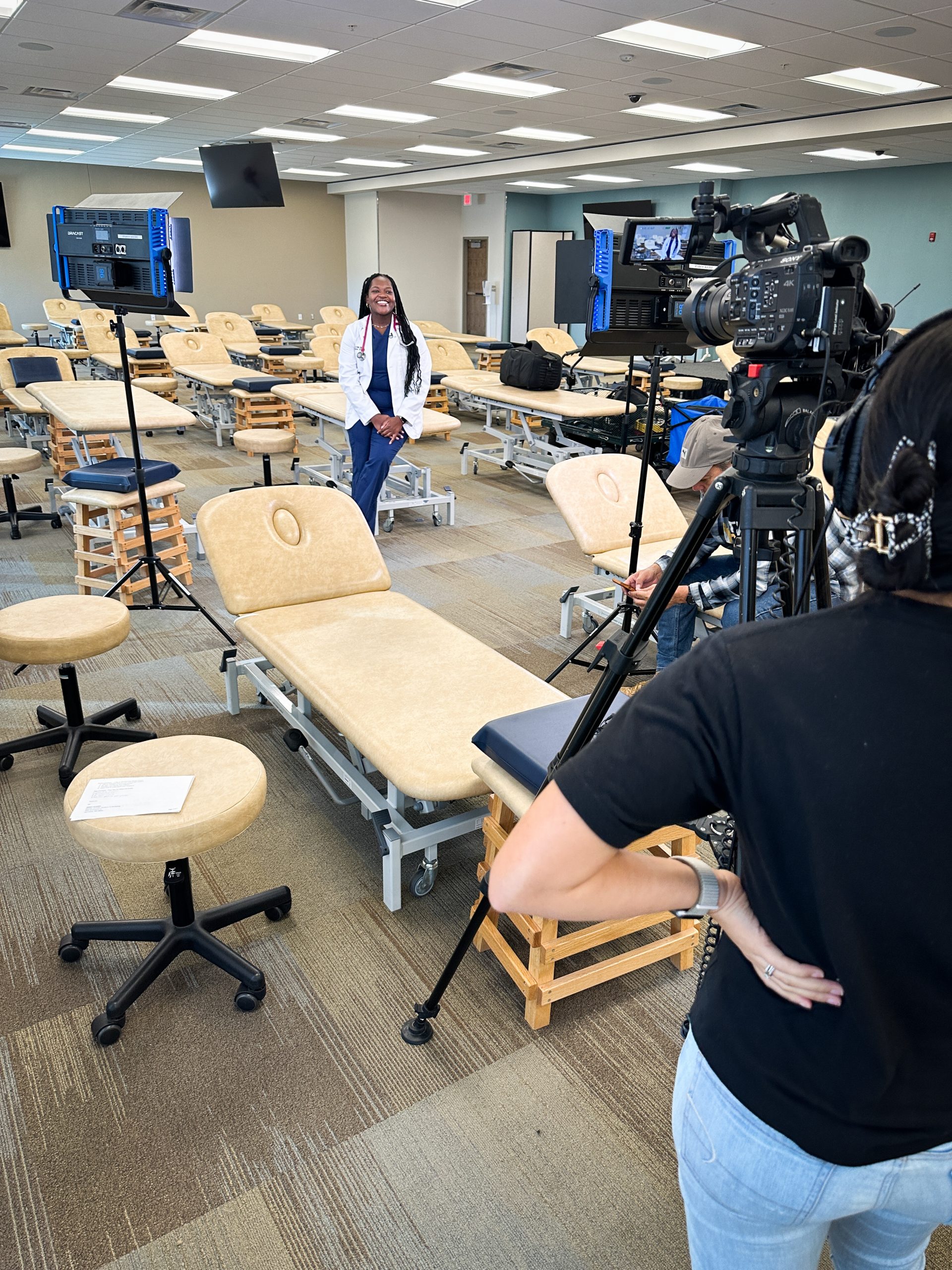
The gross anatomy lab at the Burrell College of Osteopathic Medicine (BCOM) has nearly tripled in size, going from 791 sq. ft. to 2,225 sq ft. after major renovations over the summer. The expansion will help improve medical students’ lessons and experience in clinical human anatomy.
“There is growing recognition that cadaver-based anatomy is a critical component of medical education,” says Jon Jackson, PhD, director of the gross anatomy lab. “There has also been an increasing desire from our medical students to experience detailed instruction in human anatomy and function.”
In their first year of medical school, BCOM students work with the bodies of individuals who have donated their remains after passing. This hands-on training provides students with a unique opportunity to learn about the structures of the human body and how they function. The knowledge and skills acquired through these examinations are an essential part of medical education.
Prior to the expansion, only one-fourth of a class fit into the gross anatomy lab. The tight space made curricular instruction difficult and required a complex schedule to ensure all students received enough time and training in the lab. “Now we can easily accommodate half of the entire first-year class in an experience of exploration, discovery and self-assessment,” Jackson says.
First-year medical students can now be split into two groups. While one half works with standardized patients using ultrasonography to visualize important anatomical and clinical landmarks, the other focuses on three-dimensional relationships and identification of structures in the cadaver lab.
Besides the additional square footage, sweeping changes have been to improve the student experience. Lab upgrades include a state-of-the-art scrub station for students to practice scrubbing; space for additional dissection tables to enable small group prosection-based teaching; a cadaver storage unit to support long-term storage and maintenance of teaching specimens; and new sinks, cabinetry and safety stations.
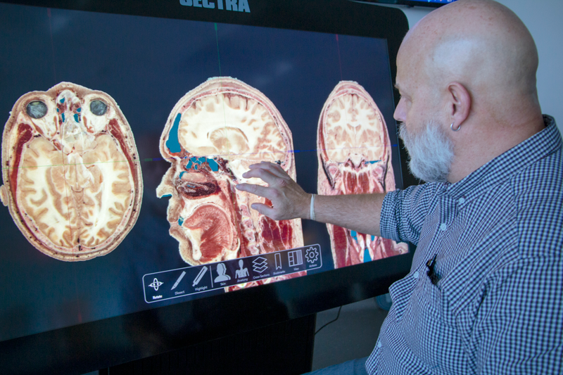
Medical students will also appreciate the lab’s technological enhancements, including multiple monitors spaced out across the room, as well as a SECTRA digital cadaver table that has high-resolution images of cadaver anatomy, histology and radiographic imaging.
Jackson says, “We are blessed to have this new, upgraded facility. It is a real pleasure to be part of the transformative experience with students as the basic science from the classroom gets integrated with the very real.”